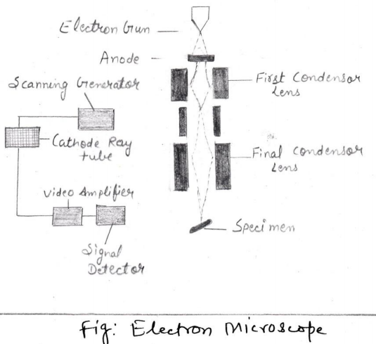
In electron microscopy, beams of electrons are used to produce images. As we already know that wavelength of electron beam is much shorter than light, resulting in much higher resolution. Generally, two types of electron microscopy is used to study the very minute objects-
1. The Transmission Electron Microscope (TEM)
We have studied in earlier classes that electrons scatter when they pass through thin sections of a specimen.
Transmitted electrons (those that do not scatter) are used in this microscopy to produce image where denser regions in specimen, scatter more electrons and appear darker.
2. The Scanning Electron Microscope(SEM)
As already discussed that electrons scatter when they pass through thin sections of a specimen. Scattered electrons produce image of thin sections in this microscopy.
Working of SEM
In SEM, the electron beam comes from a filament, made of various types of materials. For this purpose, the most commonly the Tungsten gun is used. The filament is a loop of tungsten which works as the cathode. A voltage is applied to the loop, causing it to heat or warm up. Again there is a anode, which is positive with respect to the filament, responsible for attractive forces for electrons.
It is an important phenomenon here that electrons accelerate toward the anode. They accelerate right by the anode and hit the sample through column.
Specimen preparation in Electron Microscopy
In Electron Microscopy different procedures used thanlight microscopy. In transmission electron microscopy, specimens should be cut very thin as specimens are chemically fixed and stained with electron dense material. Other preparation methods like
(i) Shadowing where coating of specimen with a thin film of a heavy metal is done.
(ii) Freeze-etching where specimen are Freezethen fracture along lines of greatest weakness (e.g. Membranes). In Scanning Electron Microscope electrons are reflected from the surface of a specimen to create image and it produces a 3-dimensional image of specimen”s surface features.


0 Comments